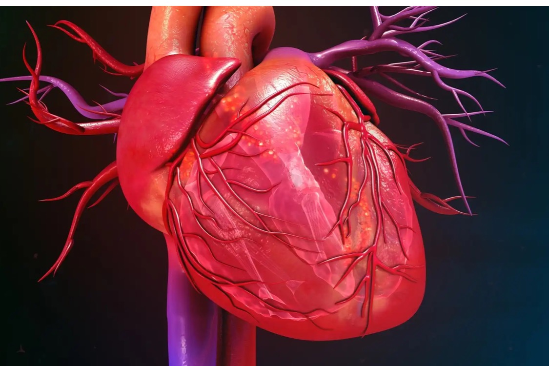
"Researchers have developed a new artificial intelligence (AI) model capable of evaluating cardiac function and identifying signs of valvular heart disease in chest X-rays. The group, sharing its work in The Lancet Digital Health, noted that this could be especially helpful in areas where qualified physicians or ultrasound technology are in short supply.[1]
The team focused on data from more than 22,000 X-rays—each associated with a corresponding echocardiogram—for its research. Their goal was to teach the AI model how the different modalities correspond with one another, using data from different sources to reduce any risk of bias. Tests from three different facilities in Japan were used for training, validating and internal testing purposes; tests from a fourth facility in Japan were used for external testing.
Overall, the team’s AI model achieved an area under the ROC curve (AUC), accuracy, sensitivity and specificity of 0.92, 86%, 82% and 86%, respectively, for the classification of left ventricular ejection fraction at a 40% cutoff. For classifying tricuspid regurgitant velocity at a 2.8 m/s cutoff, meanwhile, the model’s AUC, accuracy, sensitivity and specificity were 0.85, 75%, 83% and 73%, respectively.
The model also achieved an AUC, accuracy, sensitivity and specificity of 0.83, 73%, 79% and 72%, respectively, for the classification of aortic stenosis and an AUC, accuracy, sensitivity and specificity of 0.83, 68%, 88% and 67%, respectively, for the classification of mitral stenosis.
All of these calculations, the team added, occur “in a fraction of the time” it would take if relying on a radiologist to interpret an echocardiogram...."
Lire la suite
No echocardiography, no problem? AI evaluates cardiac function using chest X-rays
CARDIOVASCULARBUSINESS, 13/07/2023
Partagé par :
Beesens TEAM